
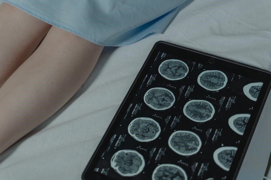
Brainwave frequency charts, often available as PDF documents, visually represent the spectrum of electrical activity in the brain.
These charts are essential tools for understanding and interpreting EEG data, aiding in neurofeedback and various clinical applications.
Analyzing brainwave patterns, ranging from delta (under 4 Hz) to gamma (up to 100 Hz), provides insights into cognitive states and neurological health.
What are Brainwaves?
Brainwaves are rhythmic patterns of electrical activity continuously generated by the synchronized firing of neurons within the brain. These oscillations, measurable using an electroencephalogram (EEG), vary in frequency and amplitude, reflecting different states of consciousness and cognitive processes.
Represented on a brainwave frequency chart, often found in PDF format, these waves are categorized into five primary types: delta, theta, alpha, beta, and gamma. The frequency is measured in Hertz (Hz), indicating cycles per second, with lower frequencies associated with deeper states like sleep and higher frequencies linked to active thinking and focused attention.
Understanding these frequencies, as detailed in available charts, is crucial for interpreting brain activity and identifying potential neurological imbalances.

Why Use a Brainwave Frequency Chart?
A brainwave frequency chart, commonly accessed as a PDF, serves as a vital reference for professionals and individuals seeking to understand brain activity. These charts provide a standardized visual guide to interpret EEG data, correlating specific frequency ranges with distinct mental states – from deep sleep to heightened concentration.
Utilizing these charts aids in identifying imbalances or anomalies in brainwave patterns, which can be indicative of neurological conditions or cognitive impairments. They are essential tools in neurofeedback and biofeedback training, allowing for targeted brainwave modulation.
Furthermore, charts facilitate research, fatigue detection, and a deeper understanding of the brain’s complex electrical language.
The Five Primary Brainwave Frequencies
Brainwave frequency charts (PDF) categorize brain activity into five main bands: delta, theta, alpha, beta, and gamma, each linked to specific states of mind.
Delta Waves (0.5 ⎻ 4 Hz)
Delta waves, ranging from 0.5 to 4 Hz, represent the slowest brainwave frequency and are prominently featured in brainwave frequency charts (PDF). These waves are dominant during deep, restorative sleep – specifically stages 3 and 4 – and are crucial for physical healing and immune system regulation.
PDF charts illustrate delta waves as having the highest amplitude, signifying strong electrical signals. They are also characteristic of infant brain activity, diminishing with age. While typically associated with unconsciousness, delta activity can sometimes appear in awake adults experiencing deep relaxation or specific abnormal brain processes, as noted in neurophysiological studies.
Understanding delta wave presence and amplitude, as visualized on a brainwave chart, is vital for assessing sleep quality and identifying potential neurological concerns.
Characteristics of Delta Waves
Delta waves, as depicted in brainwave frequency chart PDFs, are distinguished by their slow frequency – between 0.5 and 4 Hz – and remarkably high amplitude. This combination signifies a powerful, yet sluggish, electrical signal originating from the brain. PDF representations often showcase these waves as broad and sweeping, visually emphasizing their dominant presence during deep sleep.
These waves are typically most prominent over the occipital regions of the brain, though distribution can vary. They are associated with a loss of awareness of the external world and a shift towards internal processing. Brainwave charts highlight that delta activity is not merely a sign of sleep, but also a key indicator of restorative brain function.
Analyzing these characteristics via PDF charts aids in understanding sleep architecture and identifying potential neurological irregularities.
Delta Waves and Sleep Stages
Brainwave frequency chart PDFs clearly illustrate the strong correlation between delta waves and specific sleep stages. Predominantly, delta activity surges during stages 3 and 4 of Non-Rapid Eye Movement (NREM) sleep – often referred to as deep sleep or slow-wave sleep. These PDF charts visually demonstrate a significant increase in delta wave presence as one descends into deeper sleep levels.
During these stages, the brain exhibits minimal metabolic activity, allowing for crucial restorative processes. Delta waves facilitate physical healing, immune system strengthening, and memory consolidation. PDF analyses show that the proportion of time spent in delta sleep decreases with age, potentially explaining age-related changes in restorative capacity.
Understanding this relationship, as presented in brainwave charts, is vital for sleep disorder diagnosis and treatment;
Theta Waves (4 ⎻ 8 Hz)
Brainwave frequency chart PDFs depict theta waves as oscillating between 4 and 8 Hz, representing a state bridging conscious thought and deeper subconscious realms. These charts highlight theta’s prominence during drowsiness, light sleep, and profound relaxation. They are also notably present during meditative states, offering a visual representation of altered consciousness.
PDF analyses reveal that theta activity is dominant in young children, gradually decreasing with age as beta waves become more prevalent. However, adults can intentionally induce theta states through practices like meditation and deep relaxation techniques. Brainwave charts demonstrate a link between increased theta power and enhanced creativity.
These charts are invaluable for understanding the brain’s transition between wakefulness and sleep, and for exploring the potential of theta states for cognitive enhancement.
Theta Waves and Creativity
Brainwave frequency chart PDFs illustrate a strong correlation between theta wave activity and heightened creativity. These charts visually demonstrate that accessing the 4-8 Hz frequency range can unlock subconscious thought processes, fostering innovative thinking and problem-solving. Increased theta power is often observed during artistic endeavors, brainstorming sessions, and moments of inspired insight.
PDF analyses suggest that theta waves facilitate access to memories and emotions, allowing for novel connections and associations. This state is conducive to “thinking outside the box” and generating original ideas. Brainwave charts are used in neurofeedback to train individuals to intentionally increase theta activity, aiming to boost creative potential.
Understanding this link, as depicted in these charts, empowers individuals to leverage theta states for enhanced creative output.
Theta Waves and Meditation
Brainwave frequency chart PDFs consistently demonstrate a significant increase in theta wave activity during meditative states. These charts visually represent the shift from faster beta waves, associated with active thinking, to the slower theta frequencies (4-8 Hz) characteristic of deep relaxation and inner focus.
Experienced meditators often exhibit dominant theta rhythms, indicating a state of profound calmness and reduced mental chatter. PDF resources highlight that theta waves are linked to accessing subconscious material and intuitive insights, commonly experienced during meditation.
Brainwave charts are utilized in neurofeedback protocols to help individuals learn to self-regulate their brain activity, promoting deeper meditative experiences and associated benefits like stress reduction and emotional well-being.
Alpha Waves (8 ⎻ 12 Hz)
Brainwave frequency chart PDFs clearly illustrate alpha waves as prominent when individuals are in a relaxed, yet wakeful state. Typically ranging from 8 to 12 Hz, these frequencies are often observed with eyes closed and a calm mental focus. Charts demonstrate alpha activity increases during periods of quiet contemplation and reduced sensory input.
These PDF resources show alpha waves are not merely indicative of relaxation; they also play a role in cognitive performance. A moderate level of alpha activity is associated with improved attention and information processing.
Brainwave charts are valuable tools for assessing an individual’s baseline alpha rhythm and tracking changes in response to various stimuli or interventions.
Alpha Waves and Relaxation
Brainwave frequency chart PDFs consistently depict a strong correlation between alpha waves (8-12 Hz) and states of relaxation. These charts visually demonstrate increased alpha activity when an individual is calm, peaceful, and not actively processing information. Closing the eyes typically triggers a rise in alpha wave amplitude, as shown in detailed PDF representations.
The charts illustrate that alpha waves are dominant during wakeful rest, signifying a shift away from focused attention. They represent a mental state conducive to stress reduction and overall well-being.
Analyzing PDF charts helps professionals identify and promote relaxation techniques by monitoring alpha wave patterns, offering a quantifiable measure of calmness.
Alpha Waves and Cognitive Performance
Brainwave frequency chart PDFs reveal a nuanced relationship between alpha waves (8-12 Hz) and cognitive function. While often associated with relaxation, alpha activity isn’t simply a sign of inactivity. Charts demonstrate that a moderate level of alpha synchronization can actually enhance certain cognitive processes.
Specifically, PDF analyses show alpha waves play a role in integrative processing, allowing the brain to consolidate information and prepare for action. They facilitate a state of “relaxed alertness,” improving focus and problem-solving abilities.
Charts help visualize how optimal cognitive performance isn’t about eliminating alpha waves, but rather achieving a balanced brainwave state.
Beta Waves (12 ⎻ 30 Hz)
Brainwave frequency chart PDFs clearly illustrate beta waves as indicators of active mental engagement, ranging from 12 to 30 Hz. These frequencies are dominant when we are alert, attentive, and actively thinking. Charts demonstrate beta activity increases during problem-solving, decision-making, and focused concentration.
However, PDF resources also highlight that excessive beta activity can correlate with anxiety and stress. Understanding the nuances within the beta range is crucial.
Charts often detail subdivisions: low beta (15-18Hz) for quiet focus, mid beta (18-25Hz) for active thinking, and high beta (25-30Hz) potentially linked to agitation. Analyzing these distinctions is key to interpreting EEG data effectively.
Beta Waves and Active Thinking
Brainwave frequency chart PDFs consistently demonstrate a strong correlation between beta waves (12-30 Hz) and states of active thinking. When engaged in cognitive tasks – problem-solving, calculations, or focused analysis – beta activity significantly increases, as visually represented on these charts.
These PDF resources illustrate that beta waves aren’t simply present during thought, but their amplitude and specific frequency within the range (low, mid, high) can indicate the intensity of mental effort.
Charts often show beta dominance when individuals are consciously processing information, making decisions, or experiencing heightened alertness. Understanding this relationship is vital for interpreting EEG data and assessing cognitive function.
Subdivisions of Beta Waves (Low, Mid, High) ー 15Hz to 25Hz
Brainwave frequency chart PDFs detail the nuanced subdivisions within the beta range (12-30 Hz), specifically highlighting low (15-18 Hz), mid (18-25 Hz), and high beta (25Hz+) frequencies. These charts demonstrate that each subdivision correlates with distinct mental states.
PDF resources indicate low beta is associated with relaxed focus and concentration, while mid-beta signifies active, engaged thinking. High beta, as shown on these charts, often accompanies anxiety, agitation, or intense mental activity.
Analyzing these specific beta frequencies, as presented in detailed PDF guides, is crucial for precise neurofeedback training and understanding the subtleties of cognitive and emotional states.
Gamma Waves (30 ー 100 Hz)
Brainwave frequency chart PDFs illustrate gamma waves as the fastest brainwave frequency, ranging from 30 to 100 cycles per second. These charts emphasize gamma’s association with higher cognitive functions, including complex problem-solving, information processing, and conscious perception.
PDF resources detail how gamma waves are linked to binding sensory input, creating a unified experience of the world. They also play a role in learning and memory consolidation.
Detailed brainwave charts often show gamma activity increasing during focused attention and peak performance. Understanding gamma wave patterns, as presented in these PDF guides, is vital for optimizing cognitive function and neurological health.
Gamma Waves and Higher Cognitive Functions
Brainwave frequency chart PDFs consistently highlight the connection between gamma waves and advanced cognitive processes. These charts illustrate how gamma activity is prominent during tasks demanding focused attention, complex thought, and insightful problem-solving.
PDF resources explain that gamma waves facilitate the binding of sensory information, creating a cohesive and unified perception of reality. They are crucial for learning, memory formation, and conscious awareness.
Analyzing gamma wave patterns, as detailed in these PDF guides, provides insights into cognitive performance and neurological health. Increased gamma activity often correlates with heightened intelligence and efficient information processing.
Gamma Waves and Sensory Processing
Brainwave frequency chart PDFs demonstrate a strong link between gamma waves and how the brain processes sensory input. These charts illustrate that gamma activity is heightened when the brain integrates information from various senses – sight, sound, touch, and more – creating a unified perceptual experience.
PDF resources explain that gamma oscillations help synchronize neural activity across different brain regions, enabling efficient sensory binding. This synchronization is vital for recognizing patterns and making sense of the surrounding environment.
Analyzing gamma wave patterns, as shown in these PDF guides, can reveal how effectively the brain filters and prioritizes sensory information, impacting attention and awareness.
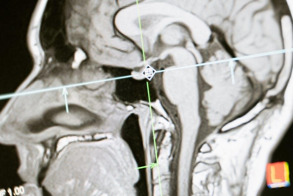
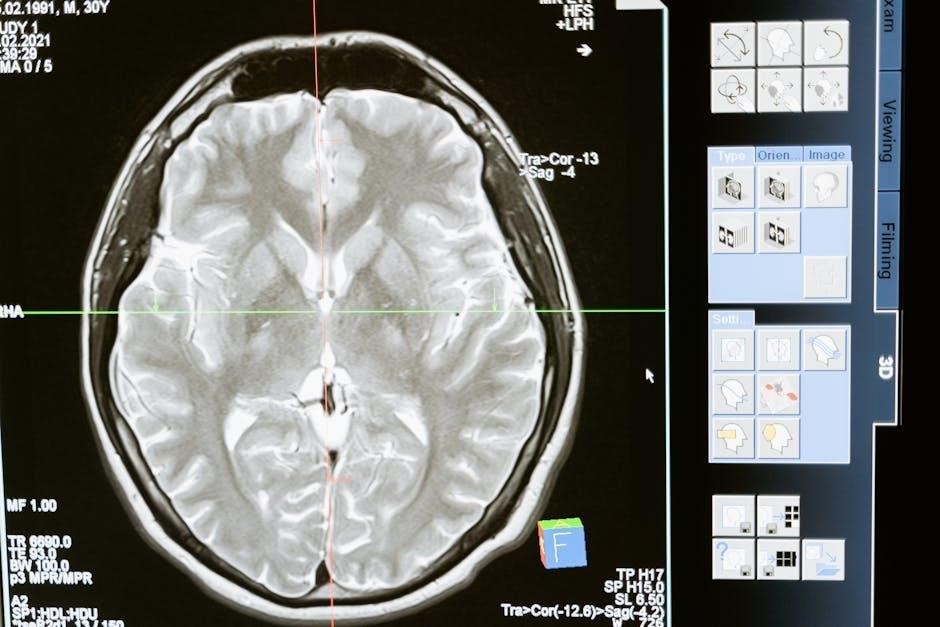
Understanding EEG and Brainwave Measurement
EEG, often visualized with brainwave frequency chart PDFs, detects electrical activity in the brain using electrodes.
These charts help quantify and interpret the recorded signals for analysis.
Quantitative Electroencephalogram (qEEG)
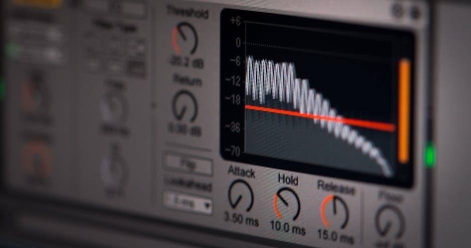
qEEG represents a sophisticated evolution of traditional EEG, employing statistical analysis and computer processing to map brainwave activity. Utilizing brainwave frequency chart PDFs as a reference, qEEG transforms raw EEG data into quantifiable metrics, often expressed as power spectral density values;
This process allows for a detailed visualization of brainwave frequencies – delta, theta, alpha, beta, and gamma – across different brain regions. The resulting maps, frequently presented in color-coded formats, highlight areas of increased or decreased activity.
qEEG is invaluable for identifying neurological patterns associated with conditions like ADHD, anxiety, and sleep disorders, offering objective data to complement clinical assessments. Software converts frequencies to magnitude (µV2) for precise analysis.
Converting Brainwave Frequencies to Magnitude (µV2)
Transforming brainwave frequencies into magnitude, measured in microvolts squared (µV2), is a crucial step in quantitative EEG (qEEG) analysis. Software applications utilize brainwave frequency chart PDFs as guides during this conversion process, allowing for standardized interpretation.
This conversion isn’t simply about identifying the frequency band (e.g., alpha, 8-12 Hz); it’s about quantifying the power within that band; Higher µV2 values indicate greater amplitude and, therefore, more prominent activity at that specific frequency.
This magnitude representation enables clinicians to compare activity across different brain regions and individuals, identifying deviations from normative databases. Analyzing these values, alongside frequency charts, provides a detailed picture of brain function.
Applications of Brainwave Frequency Analysis
Brainwave frequency chart PDFs facilitate neurofeedback, biofeedback, and fatigue detection by analyzing alpha, beta, and theta wave ratios for mood and performance insights.
Neurofeedback and Brainwave Training
Neurofeedback utilizes brainwave frequency chart PDFs as a core component, providing visual representations of a patient’s brain activity in real-time. This allows individuals to learn self-regulation techniques, shifting brainwave patterns towards desired frequencies.
By observing their brainwaves on a screen – guided by the chart’s frequency ranges – patients receive feedback (audio or visual) when their brain activity moves in the correct direction. This process strengthens neural pathways associated with focus, relaxation, or other targeted states.
PDF charts are crucial for therapists to establish baseline measurements and track progress during neurofeedback sessions, tailoring training protocols to individual needs and optimizing outcomes. They help visualize the impact of training on specific frequencies like beta for concentration.
Biofeedback and Mood Control

Biofeedback, often informed by brainwave frequency chart PDFs, extends beyond direct brainwave manipulation to encompass physiological responses linked to mood. While neurofeedback directly targets brainwaves, biofeedback uses sensors to monitor heart rate variability, muscle tension, and skin conductance – all influenced by brain activity.
PDF charts help clinicians understand the correlation between specific brainwave patterns (like increased beta activity associated with anxiety) and physiological indicators. This allows for a holistic approach to mood regulation, teaching individuals to control their bodily responses.
By learning to modulate these responses, individuals can gain greater control over their emotional states, reducing stress and improving overall well-being. Charts provide a visual guide for both therapist and patient, tracking progress and reinforcing self-regulation skills.
Fatigue Detection Using Brainwave Ratios ー (α θ)/β and α/β indexes
Brainwave frequency chart PDFs are crucial in fatigue research, particularly when analyzing ratios like (α + θ)/β and α/β. These indexes, derived from quantitative EEG (qEEG) data, offer objective measures of mental fatigue levels in professions like medicine, where on-call doctors experience significant strain.
Software utilizes these PDF-referenced charts to convert brainwave frequencies – alpha, beta, and theta – into magnitude (µV2) power. A higher (α + θ)/β ratio generally indicates increased fatigue, suggesting a shift towards slower brainwave activity. Similarly, a decreased α/β ratio points to reduced alertness.
These ratios provide a quantifiable way to assess cognitive state, aiding in fatigue detection and potentially informing strategies to mitigate the risks associated with prolonged exhaustion.
Resources for Brainwave Frequency Charts (PDF)
Brainwave frequency chart PDFs are widely available online from neurohealth associates and research publications, offering valuable references for EEG interpretation and analysis.
Where to Find Reliable Charts
Finding trustworthy brainwave frequency charts in PDF format requires careful source evaluation. NeuroHealth Associates provides resources, alongside publications on platforms like PubMed Central (PMC), offering scientifically-backed information. University websites and research institutions often host detailed charts for educational purposes.
Beware of overly simplistic or commercially driven charts lacking scientific rigor. Look for charts referencing quantitative electroencephalogram (qEEG) standards and clearly defining frequency bands (Delta, Theta, Alpha, Beta, Gamma). Reputable sources will also indicate the typical amplitude ranges associated with each frequency. Prioritize charts from peer-reviewed journals or established neurofeedback organizations to ensure accuracy and reliability.
Always cross-reference information from multiple sources.
Interpreting Brainwave Charts
Interpreting brainwave frequency charts (PDF) demands understanding that frequencies represent different mental states. Delta waves (<4 Hz) signify deep sleep, while Gamma waves (30-100 Hz) correlate with higher cognitive functions. Charts display frequencies in Hertz (Hz) and often convert them to magnitude (µV2) using software.
Analyzing ratios like (α θ)/β and α/β indexes, as explored in fatigue studies, provides insights into mental workload and exhaustion. Remember that individual variations exist; a ‘normal’ chart is a guideline, not a rigid standard. Context is crucial – consider the individual’s age, state of alertness, and any underlying neurological conditions. Professional interpretation by a trained neurofeedback practitioner is highly recommended.
Charts aid in identifying imbalances.